委託試験の特徴
- 細胞内レベルで最大40のタンパク質を同時に定量
- FFPEまたは凍結組織の分析が可能
- CLIA認定施設で定量性の高いデータを作製
- 2019年より25以上の臨床検体解析プロジェクトを完了
- インフォマティクス解析の高度なサポートを提供
- 2種類の技術(IMCまたはCODEX)から選択可能

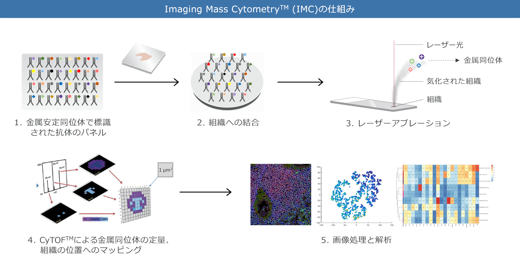
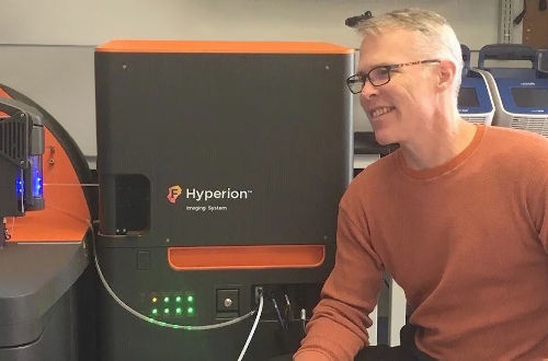
イメージングマスサイトメトリー(Imaging Mass Cytometry、IMC)は、次世代のマルチプレックス免疫化学染色であり、組織中に最大40種のタンパク質を検出できます。1: IMC用抗体は、それぞれ識別可能な金属同位体に結合されています。これにより、蛍光標識で見られる波長のオーバーラップの問題を回避できます。2: 組織切片をスライドガラスにマウントし、病理医が研究の目的に応じて分析対象の領域をマークします。一般に組織切片ごとに、1mm2領域の3カ所の分析を行います。続きて、組織をすべての抗体で同時に染色します。3: 選択した組織領域は、1µM2単位(細胞内サイズに相当)でレーザーを使用して気化することで、イオン化された金属同位体が放出されます。4: 放出された金属イオンは、特殊な質量分析技術であるCyTOFによって定量化され、各同位体の濃度が組織の解剖学的な位置にマッピングされます。5: 取得したデータは、画像の加工や分析ができます。Sirona Dx社では、IMC解析にHyperionイメージングマスサイトメーター及びMaxPar®試薬 (Fluidigm社製) を使用しています。
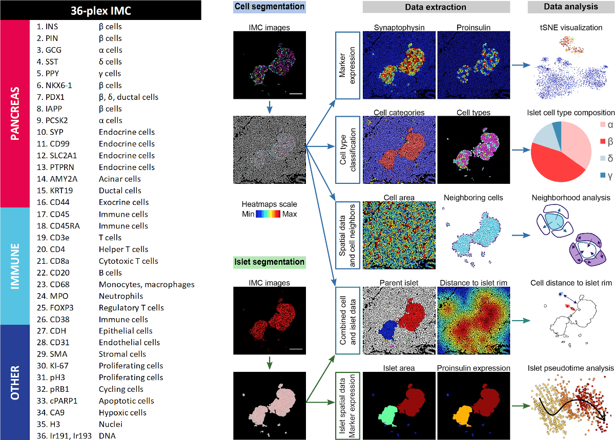
36プレックスのIMC用抗体パネルを使用して、健常人およびI型糖尿病(T1D)患者の膵臓組織における細胞種について検討しました。 使用したマーカーは左側に示しています。 図の右側は、取得したIMCデータに対して行われた膵島や細胞の解析結果の一部を示しています。 この研究により、T1Dの進行に伴うβ細胞の破壊が起こる前にβ細胞マーカーの消失と、細胞傷害性T細胞ならびにヘルパーT細胞の動員が起こるという新しい知見が得られました。 これは、IMCにより可能となる生物学的現象への洞察と、高度なインフォマティクス解析能の有用性を示します。参照文献:A Map of Human Type 1 Diabetes Progression by Imaging Mass Cytometry. Damond, N. et al. Cell Metab. 2019 Mar 5; 29(3):755-768.
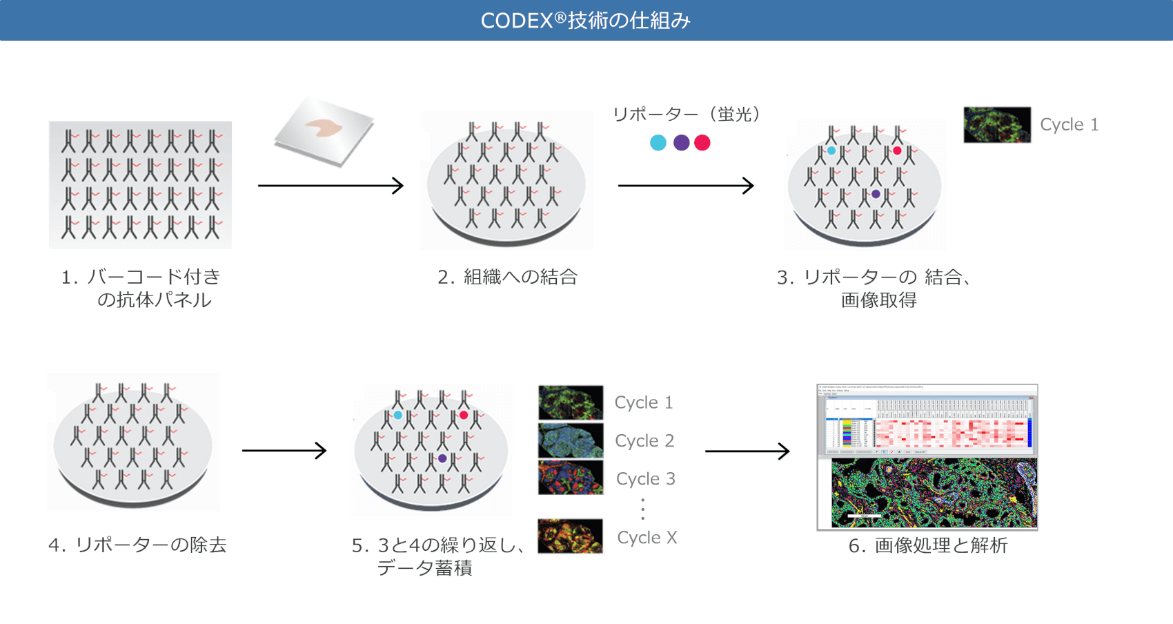
CODEX(CO-Detection by indEXing)は、マルチプレックス免疫化学染色であり、組織中に最大40種のタンパク質を検出できます。
1: CODEX用抗体には、それぞれ識別可能なDNAタグ(「CODEXバーコード」)が結合しています。2:組織切片をスライドガラスにマウントした後、すべての抗体で同時に染色されます。3:CODEXバーコードに相補的な、3つのフルオロフォア結合オリゴヌクレオチド(「リポーター」)を添加し、蛍光シグナルの画像を取得します。 波長のオーバーラップを回避するために、一回に3種類のレポーターのみでイメージングを行います。4:次に、レポーターは穏やかな条件下で除去されます。5:新しい3つのレポーターを添加し、すべての抗体が検出されるまで、抗体結合 → 画像取得 → 抗体除去のサイクルを繰り返します。 これらの操作は、専用装置により自動的に行われます。6:取得した画像データを処理して融合させた後、解析を行います。
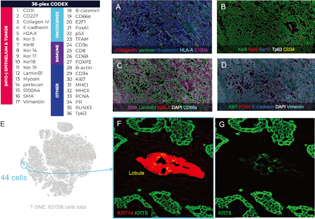
36プレックスのCODEX抗体パネルを使用して、Stage IIの腺管腺癌乳房組織中の上皮組織と、発がん遺伝子、および免疫細胞を検討しました。 使用したマーカーは左上に示しています。 A~Dは、さまざまなマーカーで染色された組織の代表的な画像を示しています。 この研究の目的の1つは、Ker14ならびにKer8を同時に発現する希少な上皮細胞種が組織に存在するか否かの確認でした。 組織サンプル中の63,056個の細胞をすべて分析し、T-SNEプロットを作成しました(E)。 これにより青色で表示された、Ker 14及びKer 8を同時に発現する小さな44個の細胞集団(総細胞集団の約0.07%)が同定されました。 画像FおよびGは、同じ実験での組織像を示しており、当該44個の細胞のKer14 / Ker8表現型を確認しています。なお、Ker8の発現は非常に弱いことが分かります。 この研究は、従来のIHC等では困難または不可能な希少細胞種の同定を、CODEX技術では可能であることを示しています。
お客様の研究にとってIMCもしくはCODEXのどちらが適切しているかは、研究の目的やお客様のご要望など、さまざまな要因によって異なります。Sirona Dx社の研究者は、最適な解析技術の選択についてもご支援いたします。
| 比較項目 | IMC | CODEX® |
| タンパク質検出 | 金属同位体結合の抗体 | DNAタグ結合の抗体 |
| 標識 | 金属同位体 | フルオロフォア |
| 最大のプレックス | 約40種類のタンパク質 | 約40種類のタンパク質 |
| 既製抗体数 | 約100種類* | 約65種類* |
| 1細胞内の解像度 | 1μM2 | 260nM2 |
| データ取得の組織面積 | 1mm2 x 3カ所が典型的 | 全面 |
| バックグラウンド | 殆どない | 多少ある |
| 抗体結合のタイミング | 全抗体を同時に | 全抗体を同時に |
| 標識のタイミング | 抗体と同時に | 3種リポーターのサイクルで |
| 抗原分解 | ない | 標識の順番は要考慮 |
| 抗体パネルの最適化 | 容易 | IMCより難しい |
| データファイルのサイズ | 小さい | 大きい |
*:新しい抗体のカスタム開発も受け付けております。
IMC及びCODEXは、それぞれに特化した抗体を使用します。既存のIMC / CODEX用抗体および抗体パネルの特異性と相互性は病理医によって検証されており、様々な組み合わせが可能です。
Sirona Dx社は、お客様から提供されたFFPEもしくは凍結組織に対して、CLIA認定施設にてIMCおよびCODEXによる解析を、委託研究サービスとして実施しています。本サービスには、試験計画、カスタム抗体の作製と検証、抗体パネルの開発と最適化、組織処理、画像取得、データ処理およびお客様の生物学的なご要望に回答するためのデータ解析が含まれます。詳細は当社までお問い合わせください。
| IMCに関する論文 | ||
| 発表年 | 詳細 | |
| 2021 | Biomarker discovery in immunotherapy-treated melanoma patients with Imaging Mass Cytometry™. Martinez-Morilla, S. et al. Clinical Cancer Research (2021): 3340. | 閲覧 |
| 2021 | ImaCytE: visual exploration of cellular micro- environments for Imaging Mass Cytometry™ data. Somarakis, A. et al. IEEE Transactions on Visualization and Computer Graphics 1 (2021): 98–110. | 閲覧 |
| 2021 | Characterisation of tumour immune microenvironment remodelling following oncogene inhibition in preclinical studies using an optimised Imaging Mass Cytometry™ workflow. Van Maldegem, F. et. al. bioRxiv (2021) *Not peer reviewed as of this time. | 閲覧 |
| 2021 | The Tumor Profiler Study: integrated, multi-omic, functional tumor profiling for clinical decision support. Irmisch, A. et al. Cancer Cell (2021): Mar 8;39(3):288-293. | 閲覧 |
| 2020 | Imaging Mass Cytometry™ and multiplatform genomics define the phenogenomic landscape of breast cancer. Ali, H.R. et al. Nature Cancer 1 (2020): 163–175. | 閲覧 |
| 2020 | Single-cell transcriptome analysis reveals disease-defining T-cell subsets in the tumor microenvironment of classic Hodgkin lymphoma. Aoki, T. et al. Cancer Discovery 10 (2020): 406–421. | 閲覧 |
| 2020 | Rare osteosarcoma cell subpopulation protein array and profiling using Imaging Mass Cytometry™ and bioinformatics analysis. Batth, I.S. et al. BMC Cancer 20 (2020): 715. | 閲覧 |
| 2020 | Single-cell mass cytometry reveals complex myeloid cell composition in active lesions of progressive multiple sclerosis. Böttcher, C. et al. Acta Neuropathologica Communications 8 (2020): 136. | 閲覧 |
| 2020 | Unraveling the complexity of the cancer microenvironment with multidimensional genomic and cytometric technologies. De Vries, N.L. et al. Frontiers in Oncology 10 (2020): 1254. | 閲覧 |
| 2020 | Oncogenic KRAS-driven metabolic reprogramming in pancreatic cancer Cells utilizes cytokines from the tumor microenvironment. Dey, P. et al. Cancer Discovery 4 (2020): 608–625. | 閲覧 |
| 2020 | A 34-marker panel for imaging mass cytometric analysis of human snap-frozen tissue. Guo, N. et al. Frontiers in Immunology 11 (2020): 1466. | 閲覧 |
| 2020 | The single-cell pathology landscape of breast cancer. Jackson, H.W. et al. Nature 578 (2020): 615–620. | 閲覧 |
| CODEX®に関する論文 | ||
| 発表年 | 詳細 | |
| 2021 | Inhibition of prostaglandin-degrading enzyme 15-PGDH rejuvenates aged muscle mass and strength. Palla et al. Science 29 Jan 2021. |
閲覧 |
| 2020 | Coordinated Cellular Neighborhoods Orchestrate Antitumoral Immunity at the Colorectal Cancer Invasive Front. Schürch et al. Cell. 2020 Sep 3;182(5):1341-1359. | 閲覧 |
| 2020 | The Society for Immunotherapy in Cancer statement on best practices for multiplex immunohistochemistry (IHC) and immunofluorescence (IF) staining and validation. Taube et al. J Immunother Cancer, 2020 8(1). | 閲覧 |
| 2020 | Overview of multiplex immunohistochemistry/immunofluorescence techniques in the era of cancer immunotherapy. Tan et al. Cancer Communications, 2020 40(4), 135-153. | 閲覧 |
| 2019 | State-of-the-art of profiling immune contexture in the era of multiplexed staining and digital analysis to study paraffin tumor tissues. Parra et al. Cancers, 2019 Feb 20;11(2):247. | 閲覧 |
| 2018 | Deep profiling of mouse splenic architecture with CODEX multiplexed imaging. Goltsev et al. Cell, 2018 174(4), 968-981. | 閲覧 |